What is Paget’s Disease of the Breast?:
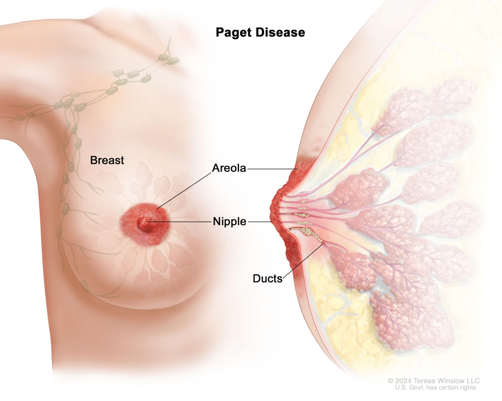
Paget’s Disease of the Breast is a rare form of breast cancer, nearly entirely affecting women. The characteristic signs of Paget’s Disease of the Breast include eczema-like flakes or scaliness of the nipple and, potentially, the areola. Discharge is also a common symptom as a result of the unique tumor cells present on the outer layers of the nipple. Though the real cause of Paget’s Disease of the Breast is unknown, most affected women have a cancer affecting their milk ducts.
Symptoms:
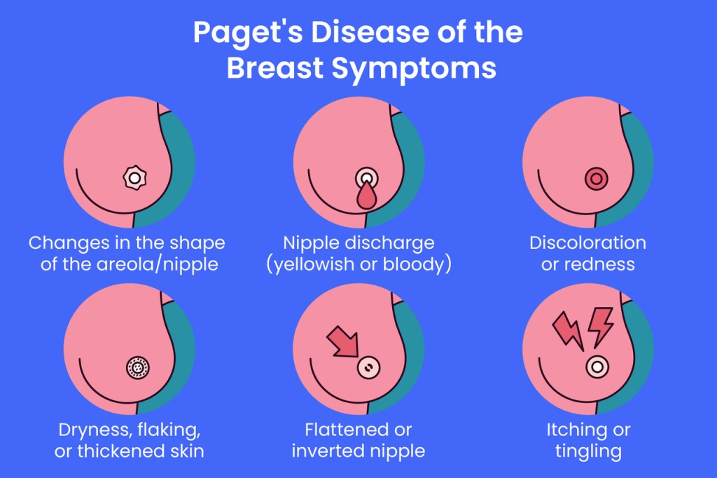
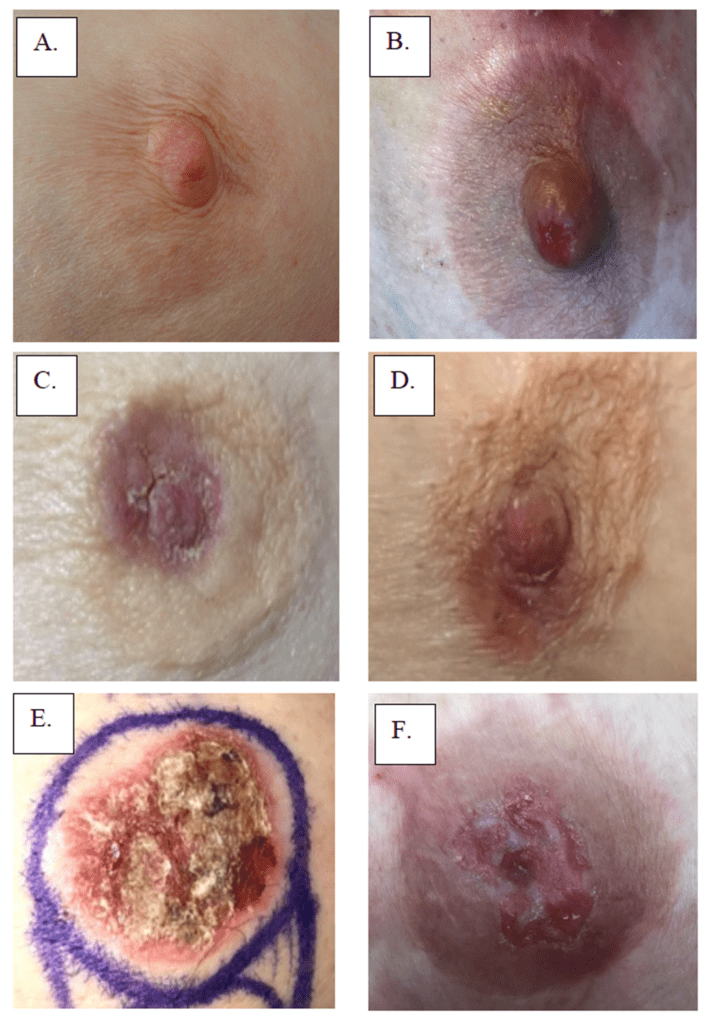
The initial changes in appearance of the nipple and surrounding areas include the development of a red, scaling, crusting, and/or abnormally thick lesions, in addition to discharge, which some affected women experience. Symptoms stemming from these lesions include itching, burning, bleeding, oozing at the site of the lesions. Pain and heightened sensitivity in the region is possible to develop over time. It is also common for symptoms to improve over time, just to worsen once more. Most often, Paget’s Disease of the Breast is unilateral, meaning it only affects one breast; however, it is possible to see a case of bilateral Paget’s Disease of the Breast, meaning it affects both breasts.
In some cases, the initial symptoms can be rather slight, being mistakenly identified as a small infection or rash. This can cause diagnosis to be delayed for months, until the itching and burning sensations drive the patients to seek medical care. Most women with Paget’s Disease of the Breast have an underlying form of cancer in the milk ducts that can sometimes be spread to other parts of the body such as the lymph nodes. Around half of those with Paget’s Disease of the Breast have found a lump beneath the nipple; however, some people with Paget’s Disease of the Breast may have other physical abnormalities such as an inverted nipple. As a result of differing conditions upon developing symptoms, the road to diagnosis and treatment vary greatly from case to case, especially depending on whether or not the individual has an underlying malignancy or cancer.
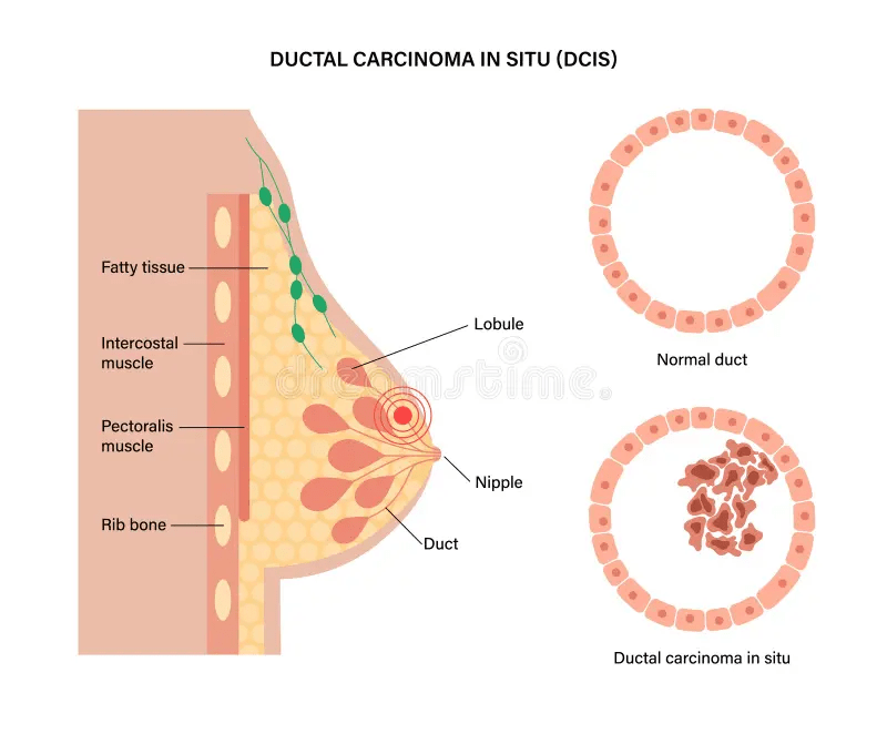
Causes:
There are two contending explanations for one’s development of Paget’s Disease of the Breast. The first theory suggests that cancerous cells from a preexisting malignancy travel via the milk ducts toward the nipples, causing Paget’s Disease of the Breast. Laboratory testing shows that the two kinds of cells share similar characteristics, as if they had a matching point of origin. Research concludes that there are cancerous cells along the lining of the milk ducts in patients with Paget’s Disease of the Breast, which are spread out to the nipple and surrounding areas, explaining the theory that Paget’s Disease of the Breast comes from preexisting cancer.
The second explanation for the occurrence of Paget’s Disease of the Breast is that it is its own disease that spreads from the outermost layer of skin on the nipple. It is suspected that this development would occur spontaneously, as the cause is still yet to be determined. This would help to explain why there are some individuals who develop Paget’s Disease of the Breast without having any underlying cancer. The root of any cancerous growth associated with Paget’s Disease of the Breast remains unknown; however, some current research takes into consideration possible genetic, immunological, and environmental factors that may lead to the development of Paget’s Disease of the Breast. Cancer itself is caused by sporadic and random changes to the structure of cells and their respective DNA, the cause is unknown. Cancer cells no longer perform their intended function and go on to pass down their abnormalities to their daughter cells, spreading the cancerous trait through the bloodstream.
Diagnosis:
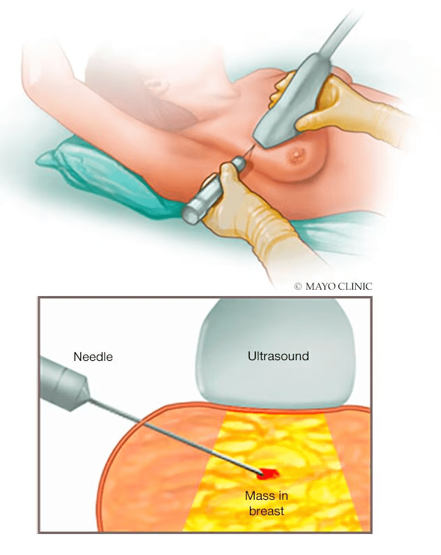
Paget’s Disease of the Breast can be diagnosed through clinical examination, identifying the characteristic physical symptoms, conducting a patient history, and a series of other specialized tests. Paget’s Disease of the Breast is commonly confused with eczema, another skin condition, causing a delay in the diagnosis. Many tests conducted include blood tests, biopsies, mammography, and/or any other imaging of the breasts or cytologic smear. A biopsy allows doctors to determine if there are any cancerous cells in the nipple tissue. During a mammogram doctors look for lumps in the breast, and if one is found, a sample of that tissue must be taken.
A mammogram is an x-ray of the breast used to identify if Paget cells are present. Mammography is also used to detect or rule out whether an underlying cancer is present. MRI may be used to create additional images of the breast, as to determine whether or not any cancer is present. Additionally, the presence of nipple discharge may also be cause for taking a sample to look for any Paget cells.
If patients have an underlying cancer, then a lymph node biopsy may have to be performed. This procedure includes collecting a sample from the axillary lymph nodes to see whether or not any invasive cancer has spread. The physician will then remove the sentinel lymph node, which is often the first lymph node cancer cells spread to.
Treatment:
The treatment of individuals with Paget’s Disease of the Breast requires a team of medical professionals, including medical oncologists, radiation oncologists, surgeons, oncology nurses and other more. Specific treatment methods vary, depending on many factors, like the extent of the primary tumor and the degree of malignancy. Elements like nature, size, and invasiveness of an underlying breast cancer, the presence or absence of metastatic disease, as well as the individual’s age and general health condition must be taken into consideration before any treatment can begin. Treatment traditionally includes the surgical removal of the breast tissue, adjacent lymph nodes, and, potentially, radical mastectomy. However, some individuals may only require a simple mastectomy, where the breast as well as the lining of the chest muscles are removed.
In the select few cases where there is no cancerous breast mass and negative mammograms, a lumpectomy may be performed. During this surgery, the nipple, areola, and a small section of breast tissue are removed. Being a conservative procedure, the physician attempts to remove as little breast tissue as necessary. This being the case, those who receive a lumpectomy must receive some follow up radiation therapy. Those with Paget’s Disease of the Breast receive therapy called adjuvant therapy, designed to go with their surgeries, preventing the cancer from coming back. This therapy can include radiation therapy, chemotherapy, and, potentially, hormone therapy.
Paget’s Disease of the Breast’s prognosis is tied to the cancer already present in the individual. However, a person with Paget’s Disease of the Breast typically does not have a prognosis that’s any worse than the underlying malignancy’s prognosis.
How You Can Make an Impact:
The cause of Paget’s Disease of the Breast remains unknown, and without proper funding and support for continued research and clinical trials to determine the root of the disease, many more women will go on to develop Paget’s Disease of the Breast. If you can, please donate here! If you are unable to donate, consider volunteering your time by raising awareness for this rare disease. If you’re interested in learning more about Paget’s Disease of the Breast, donation opportunities, or the progress being made on new treatments, visit Susan G. Komen! Susan G. Komen strives to “save lives by meeting the most critical needs in our communities and investing in breakthrough research to prevent and cure breast cancer.”
Let’s keep spreading awareness! – Lily
References:
Gadi, V. K. (2016, June 21). Paget’s Disease of the Breast – Symptoms, Causes, Treatment | NORD. NORD (National Organization for Rare Disorders); NORD. https://rarediseases.org/rare-diseases/pagets-disease-of-the-breast/

Leave a comment