What are Glioblastomas?:
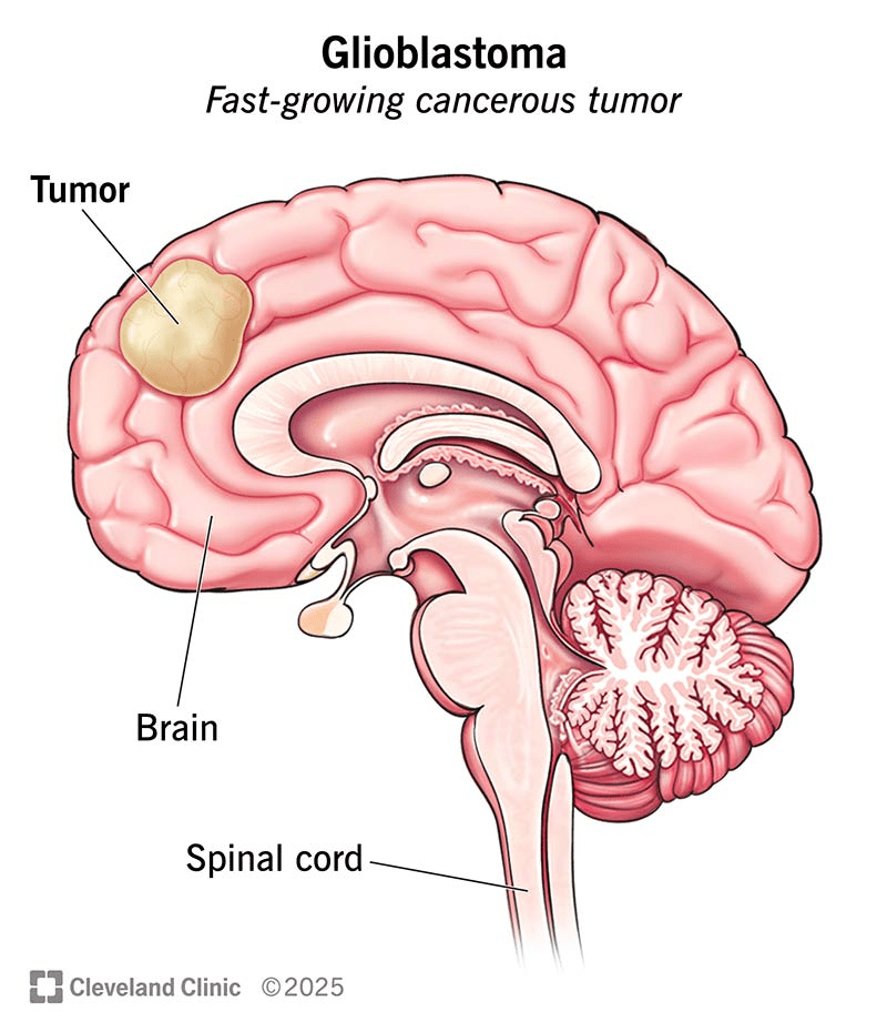
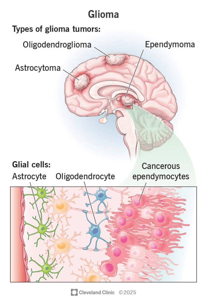
Glioblastomas are highly aggressive and malignant grade IV brain tumors that develop from glial cells, specifically a type called astrocytes. Because of this origin, they are sometimes referred to as astrocytomas. Tumors are graded from I to IV based on how fast they grow, with grade I being the slowest and grade IV being the fastest. Glioblastomas often appear as grade IV from the beginning, without progressing from a lower grade.
These tumors can form in any part of the brain but usually do not spread beyond it. Common symptoms include headaches, seizures, confusion, memory problems, muscle weakness, vision changes, difficulty with language, and changes in thinking. Glioblastomas are most often found in adults between the ages of 45 and 70, and are rare in children. Treatment usually involves a combination of surgery, chemotherapy, radiation therapy, and therapy using alternating electric fields. On average, patients who receive this combined treatment live about 14.6 months.
Symptoms:
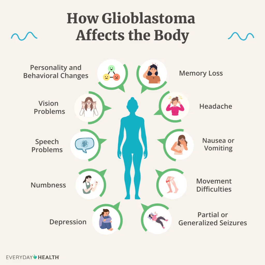
Patients with glioblastoma experience two main types of symptoms: generalized and focal. Generalized symptoms are common across many brain tumors and include headaches, seizures, nausea, vomiting, memory loss and reduced overall function. Focal symptoms depend on the tumor’s size and where it is located in the brain. For instance, a tumor in the area responsible for language may cause difficulty with speaking or understanding speech. Other focal symptoms can include seizures, muscle weakness, sensory loss and vision problems.
Tumors can also lead to brain swelling by taking up space and pressing on nearby areas. This pressure can result in headaches, nausea and vomiting. Glioblastoma grows quickly and often invades nearby brain tissue, making it a particularly aggressive form of brain cancer.
Causes:
The exact cause of glioblastoma remains unknown. However, certain factors may increase the risk of developing it. One known risk factor is previous exposure to ionizing radiation therapy, which uses high energy waves or particles to destroy cancer cells but can also harm healthy cells and potentially lead to new cancers. Other possible risk factors include working in industries such as synthetic rubber manufacturing or petroleum refining, and exposure to substances like vinyl chloride or pesticides. It is important to understand that many people diagnosed with glioblastoma have no known risk factors, and individuals with these risk factors may never develop the disease. There is no confirmed link between these factors and glioblastoma, and more research is needed.
Diagnosis:
When glioblastoma is suspected, the patient should first receive a full physical and neurological exam. Neurological exams help evaluate muscle strength and sensory responses. If symptoms suggest glioblastoma, the next step is brain imaging using contrast-enhanced magnetic resonance imaging, or MRI. This imaging method uses a strong magnetic field and radio waves to create detailed images of the brain. A contrast dye is used to make it easier to see tumors by highlighting differences between normal and abnormal tissue. While MRI can strongly suggest the presence of glioblastoma, a definitive diagnosis requires a biopsy. A biopsy involves removing a tissue sample, which is then examined to confirm if it is glioblastoma.
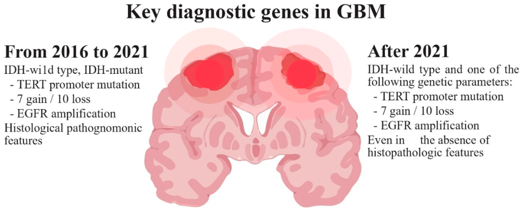
Several clinical tests are used to evaluate disease progression and treatment success. These include checking for IDH gene mutations, assessing the Karnofsky Performance Status, and determining MGMT enzyme status. Patients with IDH mutations typically have tumors that grow more slowly and respond better to treatment. The Karnofsky score measures how well a person can function in daily life. A score of 100 means the patient is fully independent, while a score of 50 means the patient needs significant help. Higher scores are linked to better treatment outcomes. MGMT is an enzyme that helps repair DNA. Its activity can influence how effective chemotherapy is. Patients with the normal version of this enzyme usually respond more positively to treatment.
Treatment:
The treatment of glioblastoma involves a team of medical professionals working together. Each specialist has a specific role. Neuro-oncologists focus on diagnosis and medical treatment, neurosurgeons perform tumor removal, radiation oncologists provide radiation therapy, nurses offer care and support, social workers address social and emotional needs, pathologists analyze tissue samples, and neuroradiologists interpret brain scans. Treatment usually includes a combination of surgery, radiation, chemotherapy, and electric field therapy.
Surgical removal of the tumor is typically the first step. The goal is to take out as much of the tumor as possible while avoiding lasting damage to the brain. Techniques to improve outcomes include awake craniotomy, fluorescent dyes, intraoperative MRI, and stereotactic guidance. Awake craniotomy allows the patient to remain conscious and communicate with the surgical team, which helps avoid impairing important functions such as speech. Fluorescent dyes highlight tumor tissue, making it easier to remove. Intraoperative MRI provides real-time imaging during surgery, and stereotactic guidance allows precise targeting of the tumor. Even after what appears to be complete removal, microscopic tumor cells remain, meaning surgery alone is not a cure.
Radiation therapy follows surgery and works by damaging the DNA of tumor cells to slow or stop their growth. Normal brain cells are also affected, which can lead to side effects such as fatigue, hair loss, appetite changes, and skin problems. Because of these side effects, radiation is only given for a limited time.
Chemotherapy can be used alongside and after radiation. Temozolomide is the most effective and widely used drug for glioblastoma. Other options approved for treatment include bevacizumab, which reduces blood supply to the tumor, and Gliadel wafers, which deliver chemotherapy directly to the brain at the tumor site.
Electric field therapy is another option, often used with chemotherapy. It is not used during radiation. This treatment involves placing electrodes on the shaved scalp. These generate alternating electric fields that interfere with cancer cell division. The treatment must be used consistently to see benefits and has shown only modest results in clinical use.
How You Can Make an Impact:
Without proper research, funding, and support for continued studies and clinical trials to determine possible cures, legitimate medicines for the disease, or preventative treatment, many more people will go on to develop glioblastomas. If you can, please donate here! If you are unable to donate, consider volunteering your time by raising awareness for this rare disease. If you’re interested in learning more about glioblastomas, donation opportunities, or the progress being made on potential treatments, visit the Glioblastoma Foundation. The Glioblastoma Foundation strives to “galvanize glioblastoma drug development.”
Let’s keep spreading awareness! – Lily
References:
Tran, T., & Bruce, J. N. (2023, November 17). Glioblastoma – Symptoms, Causes, Treatment | NORD. NORD (National Organization for Rare Disorders); NORD. https://rarediseases.org/rare-diseases/glioblastoma-multiforme/

Leave a comment